

Điều Trị Đau Clinic
Xua tan những cơn đau - All for Pain relief !

Điều Trị Đau .com được Bs Mai Trung Dũng sáng lập và phát triển dưới sự bảo trợ của Bệnh viện Quân y 354, là website hàng đầu Việt Nam về Đau và Điều trị đau cơ xương khớp, cột sống, thần kinh; website đã từng được Bộ Y tế trao GIẢI NHÌ trong Hội thao sáng tạo tuổi trẻ ngành y tế KV Hà Nội năm 2009, và được Bộ Quốc phòng tặng GIẢI NHẤT Giải thưởng tuổi trẻ sáng tạo trong Quân đội năm 2010.
ĐIỀU TRỊ ĐAU SHOP


3.500.000₫
-6%
Original price was: 3.700.000₫.3.490.000₫Current price is: 3.490.000₫.

-12%
Original price was: 2.600.000₫.2.290.000₫Current price is: 2.290.000₫.
-5%
Original price was: 11.550.000₫.10.990.000₫Current price is: 10.990.000₫.
-2%
Original price was: 2.700.000₫.2.650.000₫Current price is: 2.650.000₫.
-9%
Bán chạy

Original price was: 3.600.000₫.3.290.000₫Current price is: 3.290.000₫.

-9%

Original price was: 11.490.000₫.10.490.000₫Current price is: 10.490.000₫.
800.000₫
ĐIỀU TRỊ ĐAU CLINIC
Khám và điều trị bệnh Cơ Xương Khớp - Cột sống - Thần kinh bằng Vật lý trị liệu, Phục hồi chức năng, Châm cứu YHCT
Địa chỉ: Số nhà G11 Ngõ 28, Xuân La, Tây Hồ, Hà Nội
Hotline: 084 354 8686
Thời gian làm việc: Thứ 2-Thứ 6, từ 17h-20h;
Thứ 7 & Chủ Nhật từ 9h-12h và từ 15h-18h
Đặt lịch hẹn khám
MỘT SỐ PHƯƠNG PHÁP ĐIỀU TRỊ
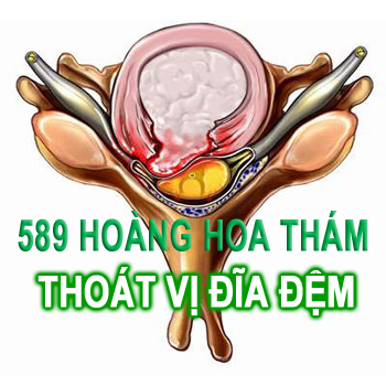
THOÁT VỊ ĐĨA ĐỆM, THOÁI HÓA CỘT SỐNG, CƠ XƯƠNG KHỚP
ĐƯỢC ĐIỀU TRỊ ĐAU CLINIC ÁP DỤNG

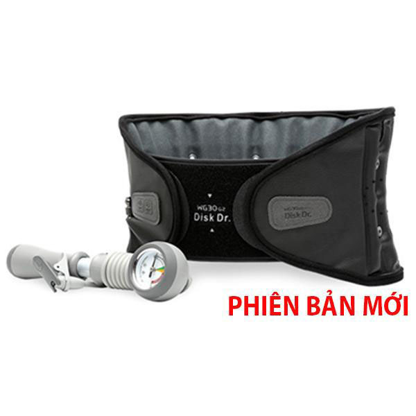
KÉO GIÃN CỘT SỐNGTác dụng: Giảm chèn ép thần kinh, giảm đau, giãn cơCơ chế: Đây là phương pháp bảo tồn duy nhất có thể tác động vào căn nguyên gây bệnh, làm giảm áp lực trong đĩa đệm giúp làm thu nhỏ khối thoát vị, mở rộng lỗ tiếp hợp làm giảm sự chèn ép thần kinh, giải quyết tận gốc của vấn đề, giúp giảm đau nhanh chóng và bền vững.
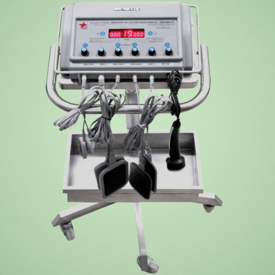
LIỆU PHÁP ĐIỆN CHÂMTác dụng: Theo YHCT, châm cứu làm thông kinh hoạt lạc, chỉ thống. Còn theo YHHĐ, điện châm làm giảm đau.Cơ chế: Châm cứu là một kích thích gây ra một cung phản xạ mới có tác dụng ức chế và phá vỡ cung phản xạ đau do thoái hóa hoặc thoát vị đĩa đệm cột sống, tăng tiết mooc-phin nội sinh tạo cảm giác dễ chịu, thư giãn. Sử dụng dòng xung của máy điện châm với kiểu xung, tần số và biến điệu phù hợp với từng bệnh nhân để mang lại hiệu quả giảm đau cao nhất.


ĐIỆN XUNG TRỊ LIỆUTác dụng: Giảm đau, giảm co thắt cơ.Cơ chế: Điện xung tạo ra xung động thần kinh có tác dụng cắt đứt đường dẫn truyền đau lên não ở tủy sống làm cho não không nhận được tín hiệu đau giúp ta không còn thấy đau nữa. Điện xung cũng giúp tăng tiết mooc-phin nội sinh giúp giảm đau, thư giãn. Điện xung còn làm giãn cơ cắt đứt vòng xoán bệnh lý Đau - Co thắt cơ.
SIÊU ÂM ĐIỀU TRỊTác dụng: giảm đau, giảm co thắt cơ.Tác dụng đầu tiên của siêu âm trong tổ chức là tác dụng cơ học, do sự lan truyền của sóng siêu âm gây nên những thay đổi áp lực tương ứng với tần số siêu âm, tạo nên hiện tượng gọi là “xoa bóp vi thể” gây ra các tác dụng sinh học làm giãn cơ rất tốt đối với các cơ đang co thắt từ đó cắt đứt vòng xoắn bệnh lý trong đau, làm mềm các tổ chức liên kết (mềm sẹo)


ĐIỆN PHÂN DẪN THUỐCTác dụng: Chống viêm, giảm đau.Cơ chế: Sử dụng dòng điện 1 chiều đều để đẩy các ion thuốc chữa bệnh vào tổ chức cơ thể. Thuốc hay dùng là các thuốc chống viêm, với phương pháp này chỉ 1 lượng nhỏ thuốc cũng có tác dụng rất lớn, và hầu như không có tác dụng phụ như sử dụng thuốc uống hoặc tiêm.
TÚI CHƯỜM THẢO DƯỢCTác dụng: giảm đau, giảm co thắt cơ.Cơ chế: Bản thân nhiệt có tác dụng giảm đau tức thì do ức chế cảm giác đau, tăng hấp thụ các chất gây đau ở tổ chức cơ thể, giãn mạch xung huyết tăng cường tuần hoàn dinh dưỡng cho tổ chức. Trong điều trị túi chườm thảo dược, sử dụng nhiệt còn để tăng cường khả năng thẩm thấu của thuốc đông y vào các tổ chức bệnh.

Ý KIẾN KHÁCH HÀNG

NSƯT Anh Thái
Tôi thường xuyên bị đau lưng do thoái hóa cột sống, nhờ điều trị châm cứu, kéo giãn tại PK Điều Trị Đau mà bây giờ không còn đau nữa, cám ơn các bác sĩ và y tá.

Đỗ Ngọc Huyền
Mỗi lần đến phòng khám Điều Trị Đau, mình được phục vụ rất chu đáo và không phải chờ đợi để được khám và kết quả lâu. Mình rất thích cách làm việc đó

Mai Khánh Chi
Mình bị thoát vị đĩa đệm, đã điều trị nhiều nơi không đỡ, bác sĩ bảo có thể mình phải đi mổ, mình rất hoang mang và lo sợ. Nhưng sau khi được các bác sĩ PK Điều Trị Đau điều trị điện châm, kéo giãn, mình đã hết đau không cần phải mổ nữa.

Liesna
Mình là người Indonesia nhưng là khách “ruột” của phòng khám, mỗi khi thấy đau lưng là mình hay qua đó để được điều trị. Nói chung là ok lắm từ thái độ phục vụ tới cách làm việc của đội ngũ nhân viên ở đó
ĐIỀU TRỊ ĐAU BOOK

Đề tài Tiếng Việt
Những tiến bộ của công nghệ terahertz trong khoa học thần kinh
Bản tóm tắt Sóng Terahertz (THz) nằm trong phạm vi giữa vùng vi sóng và hồng ngoại trong phổ điện từ. Công nghệ THz đã được chứng minh là có [...]
Đọc tiếp
03
Th5
Th5

Bệnh cơ xương khớp
Sức khỏe sinh sản của các Bệnh nhân Viêm khớp hệ thống
Sức khỏe sinh sản của các bệnh nhân viêm khớp hệ thống, một vấn đề rất nhân văn, nhưng chưa được quan tâm đầy đủ
Đọc tiếp
11
Th4
Th4

Bệnh cơ xương khớp
Cập nhật chẩn đoán và điều trị Gút không điển hình
TÓM TẮT Bên cạnh các biểu hiện viêm khớp điển hình, bệnh gút có rất nhiều thể không điển hình. Điều đó gây chẩn đoán nhầm và điều trị sai [...]
Đọc tiếp
10
Th4
Th4
C
Calcium Sandoz Injectable
Calcium Sandoz Injectable! Khi dùng liệu pháp canxi liều cao cần phải kiểm tra chặt chẽ canxi huyết và canxi niệu, nhất là ở trẻ em và bệnh nhân đang [...]
Đọc tiếpC
Calcium Sandoz Forte
Calcium Sandoz Forte! Trường hợp canxi niệu tăng nhẹ (trên 300 mg hoặc 7,5 mmol/24 giờ), suy thận vừa hoặc nhẹ hoặc tiền sử sỏi canxi, nên tăng cường theo [...]
Đọc tiếpC
Calcium Corbiere Vitamines
Các cycline: Làm giảm hấp thu các cycline ở đường tiêu hóa, do đó nên uống 2 loại thuốc cách nhau trên 2 giờ. Digitalis: Nguy cơ gây rối loạn [...]
Đọc tiếpC
Celestamine
Celestamine! Phối hợp bétaméthasone và dexchlorphéniramine maléate cho phép giảm liều corticoide mà vẫn thu được hiệu quả tương tự khi chỉ dùng riêng corticoide đó với liều cao hơn.
Đọc tiếp